Tests to examine cells and genes

Standard pathology tests
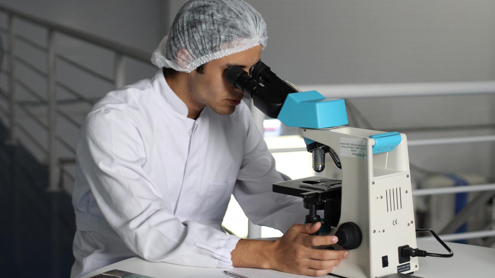
If you have a biopsy, the samples will be examined under a microscope. The pathologist will look at the size, shape and number of cells to see if they contain cancer, to diagnose the type of cancer and to give more information about the cancer.
Immunophenotyping
Immunophenotyping is a test that checks what kind of proteins or markers are on the surface of cancer cells. Cancer can produce particular proteins or make cells over-produce or stop producing particular proteins.
Knowing about these changes can help your doctor:
- Diagnose what type of cancer you have. For example, different types of leukaemia and lymphoma.
- Find out how your cancer might develop. For example, some types of proteins help the cancer to grow faster.
- Plan the best treatment for you. Some targeted therapy treatments can attack or attach to certain proteins or markers to help treat your cancer.
Immunohistochemistry and flow cytometry are examples of immunophenotyping tests. In these tests, cells are treated with antibodies in the laboratory. The antibodies stick to certain proteins on the cells. The cells are then examined to see which ones the antibodies have stuck to, helping to identify the proteins on the surface of the cells.
Cytogenetic tests
Cytogenetic tests look at chromosomes (which contain genetic information) to see if there are any abnormal changes in them.
Changes in chromosomes can help to diagnose some types of cancer, decide on the best treatment or see how well a treatment is working. For example, there may be targeted therapies that can use the genetic change to attack the cancer.
Cytogenetic tests are done in a laboratory and can use tissue, blood or bone marrow samples.
Ways of looking at the cells
Under a microscope: The cells are examined to see if there are any missing or extra chromosomes or any damage or other abnormal changes to chromosomes – for example, with some types of blood cancer translocation happens. This is when DNA from one chromosome attaches to another chromosome. An example of this type of change is the Philadelphia chromosome.
Using a dye: Fluorescent-in-situ hybridisation (FISH) uses fluorescent dyes that attach to parts of particular chromosomes. The dyes will glow (fluoresce) under ultraviolet light, to help the pathologist identify any gene changes in the cells.
FISH is helpful in finding chromosome changes that are as too small to be seen with usual cytogenetic testing. A FISH test can help your doctor to predict how your cancer might respond to a particular treatment, so they can recommend the best option for you.
Getting cytogenetic test results
In order to do some types of cytogenetic tests, scientists need to grow cells in the laboratory from the blood, bone marrow or tissue samples. This means it can take 2-4 weeks to get results of some cytogenetic tests. Sometimes the samples will need to be sent away to specialist labs, which takes longer.
For more information
Phone
1800 200 700


